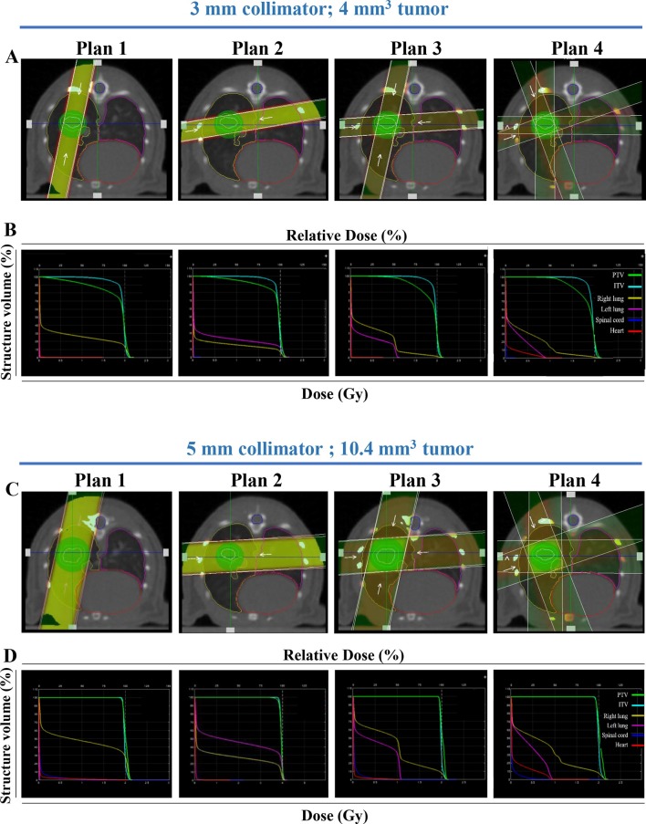
Figure 3.
Volume-dependent radiation treatment planning. Representative beam setup in axial plane for four radiation plans: parallel-opposed, equally weighted single angle, static crossing a single lung (Plan 1), crossing both lungs (Plan 2), 2-angle parallel-opposed static (Plan 3), or two equally weighted arc (Plan 4) beams for a H1299 tumor model. Two tumors volumes are shown: volume 1 (4 mm3) with the 3 mm collimator (A) and volume 2 (10.4 mm3) with the 5 mm collimator (C), with their corresponding dose–volume histograms (B, D respectively). PTV, ITV, right lung, left lung, spinal cord and heart are indicated in green, light blue, yellow, pink, dark blue and red, respectively. ITV, internal target volume; PTV, planning targetvolume.

