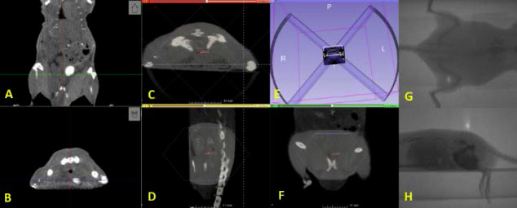
Figure 3. .
(A) Coronal and (B) axial CBCT slices showing the bladder filled with contrast. (C–F) Representative dual-arc RT plan delivering 12 Gy to the prostate using a 2 cm collimator. Portal images show (G) anteroposterior and (H) lateral beams for animal treatment position verification. RT, radiation therapy.

