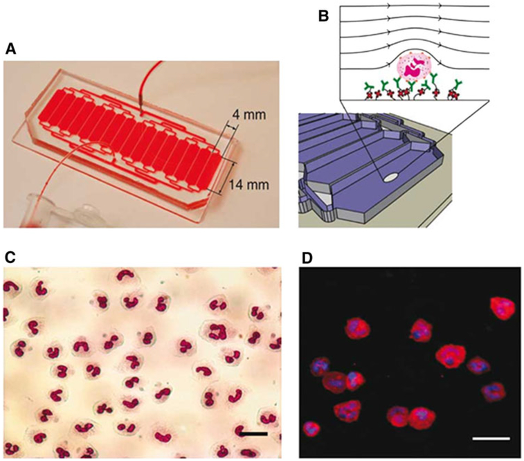
Fig. 4.
Microfluidic chip design demonstrating small size (a) and the chip surface depiction(b) with biotinylated CD66b antibodies (green) bound to neutravidin molecules (red) linked to the surface of the device. Demonstration of the microfluidic chip cell yield purity by Wright–Giemsa (c) or immunofluorescence using neutrophil cell surface markers (d) (scale bar 25 lm) (figure adapted from Kotz K et al. [42])

