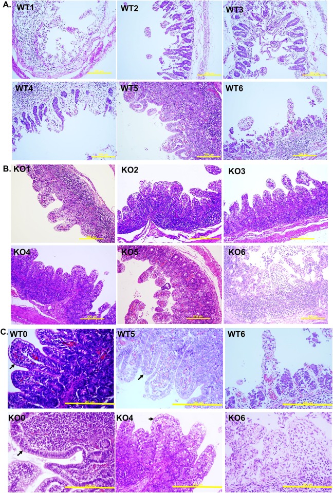
Fig 8. Pathological inspection of piglets’ intestine at the middle jejunum by H/E staining.
Panels A. and B. indicate wild-type and knockout piglets, respectively, after PEDV oral inoculation. C. The samples from control, the best and the worst pathological responded piglets, which are no nv-PEDV-inoculated, survival and dead, respectively, at 72 hpi. The upper panels (WT0, WT5 and WT6) are samples from wild type piglets and the down panels (KO0, KO4 and KO6 are samples from CMAH KO piglets. Arrows indicate epithelial cells and the yellow bars indicate 200 μm.

