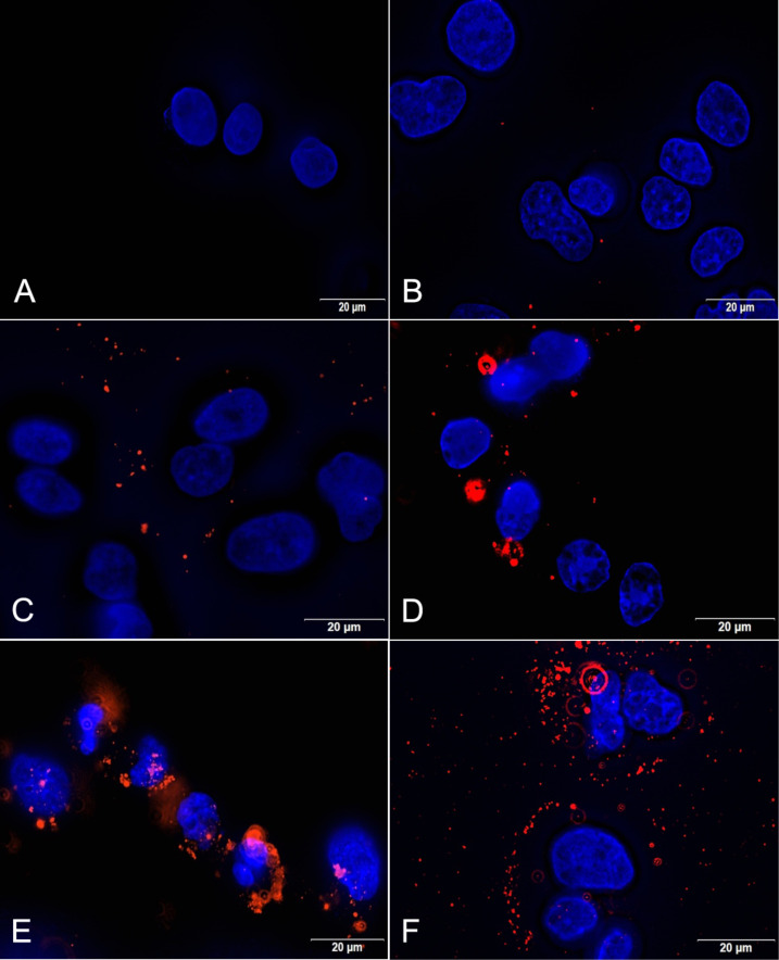
Figure 8.
Visualization of the cell interaction of PLGA NPs after 6 h with HepG2 cells in the absence (C) or presence (D–F) of a preformed protein corona. Cell nuclei were stained with DAPI (blue) and Lumogen® Red was detected through its autofluorescence (red). A: untreated cells; B: unformulated Lumogen® Red; C: PLGA NPs; D: PLGA-500-HS-NP; E: PLGA-5-HS-NP; F: PLGA-500-FBS-NP. Abbreviations: fetal bovine serum (FBS), human serum (HS).

