Figure 4.
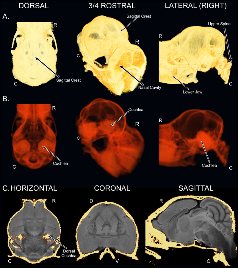
CT-based images of the mustached bat skull and registration to the T2w 3D-RARE. (A) 3D renderings of the dorsal (left), three-quarter view rostral (middle), and right hemispheric lateral (right) views of the mustached bat skull. (B) 3D renderings of the mustached bat skull from the same perspectives as in “A” except that the radiopacity has been digitally reduced using the AlphaScale functionality of AMIRA 5.4.5. (C) Horizontal (left; slice 56/126, 0.000 mm), coronal (middle; slice 166/250, 0.000 mm), and sagittal (right; slice 95/192, 0.000 mm) co-registration of the CT images of the mustached bat skull and the brain-extracted T2w 3D-RARE images of the mustached bat brain. The bilateral bony structure located within the horizontal slice is the dorsal-most peak of the cochlea.

