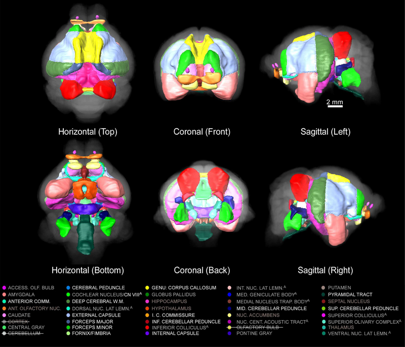
Figure 7.
Three-dimensional rendering of subcerebellar and subcortical delineations registered to the mustached bat brain (T2w 3D-RARE) from the top-horizontal (top left), bottom-horizontal (bottom left), anterior-coronal (middle top), posterior-coronal (middle bottom), left-sagittal (top right), and right-sagittal (bottom right) perspectives. The cerebellum, the cortex, and the olfactory bulb are excluded from the delineated structures shown here and in the color codes provided below to avoid obscuring subcerebellar and subcortical structures.

