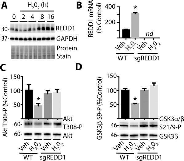
Figure 5.

ROS promotes REDD1-dependent inhibition of Akt/GSK3 phosphorylation. R28 WT and REDD1 KO (sgREDD1) cells were exposed to culture medium containing 1 mmol/L H2O2 for 0 to 24 hours. (A) REDD1 and GAPDH protein expression were assessed via Western blotting. Protein loading was evaluated by protein stain. Protein molecular mass (kDa) is indicated at left of blots. (B) REDD1 mRNA was evaluated by qPCR 24 hours after addition of either H2O2 or Veh to culture medium. Akt phosphorylation at Thr308 (C) and GSK3α/β phosphorylation at Ser21/9 (D) were assessed via Western blotting 24 hours after addition of H2O2 or Veh. Results are representative for three experiments. Within each experiment, two to three independent samples were analyzed. Values are means + SEM. *P < 0.05 versus Veh; nd, not detected.
