. 2019 May 16;8:e46740. doi: 10.7554/eLife.46740

Figure 4. A complex of several TMED proteins facilitates GFP-PrP* degradation.

(A) Wild type (WT) or TMED10 knockout (KO) cells expressing GFP-PrP* were assayed for thapsigargin-stimulated extracellular Nb uptake as in Figure 3D. In addition, the KO cells were transiently transfected with HA-tagged TMED10 lacking its coiled-coil domain (∆CC) and the transfected cells were analyzed in parallel. (B) RFP-tagged wild type (WT) TMED10, or constructs lacking the GOLD domain (residues 41–129; ∆GOLD) or coiled-coil domain (residues 130–183; ∆CC) were transiently transfected into ∆TMED10 cells inducibly expressing GFP-PrP*. GFP-PrP* was either left uninduced or induced for 48 hr prior to analysis. GFP-PrP* was immunoprecipitated using sepharose-conjugated anti-GFP Nb and analyzed by immunoblotting for GFP and RFP relative to input lysates. All input samples are from the same blot and exposure, with the vertical line indicating where intervening lanes were removed. All of the IP samples are also from the same blot and exposure. (C) Wild type or TMED10 knockout cells were transiently transfected with RFP-tagged WT or ∆CC TMED10. Two days post-transfection, cells containing moderate levels of RFP were isolated by flow cytometry, lysed, and immunoblotted against TMED2.
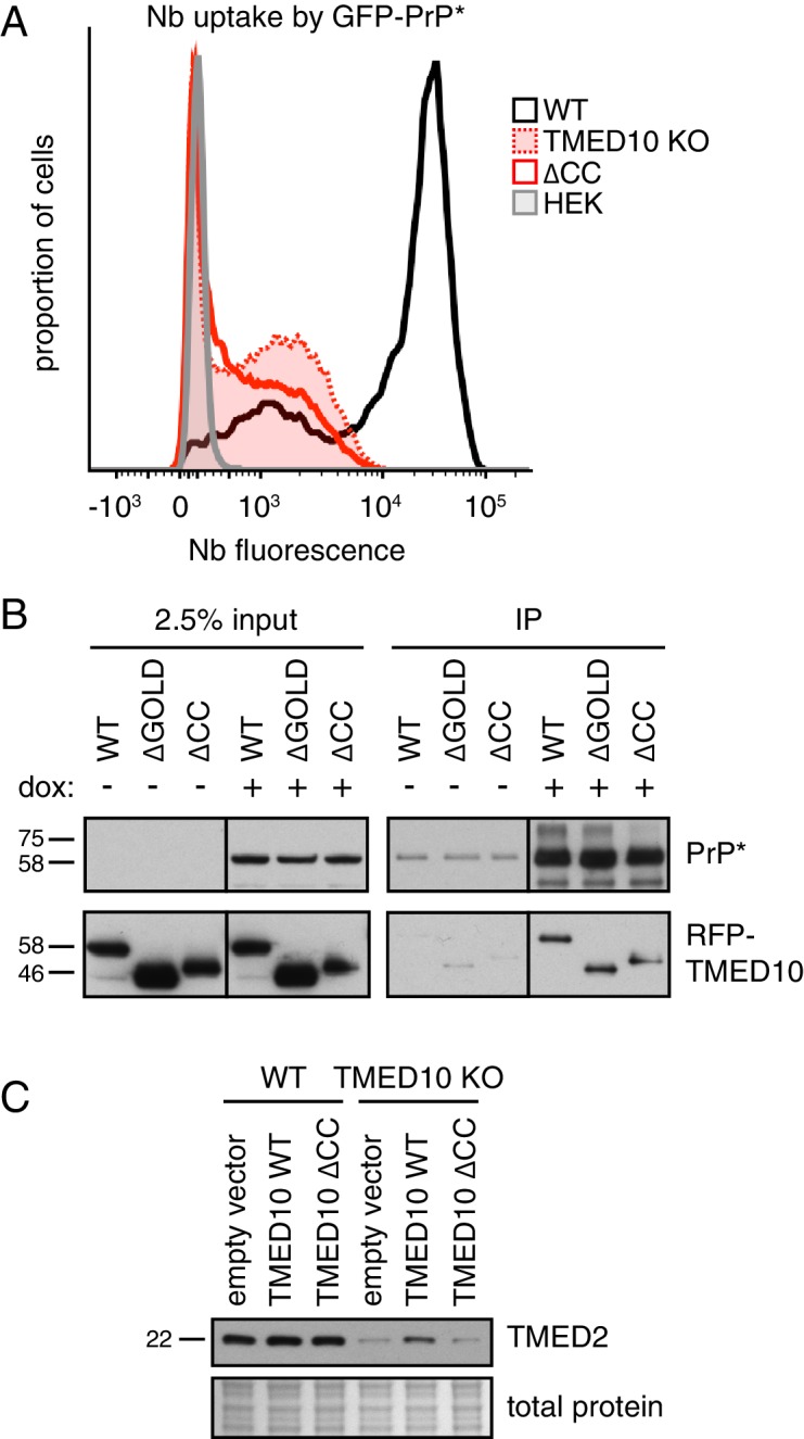
Figure 4.

Figure 4—figure supplement 1. Additional characterization of TMED family members.

Figure 4—figure supplement 1.

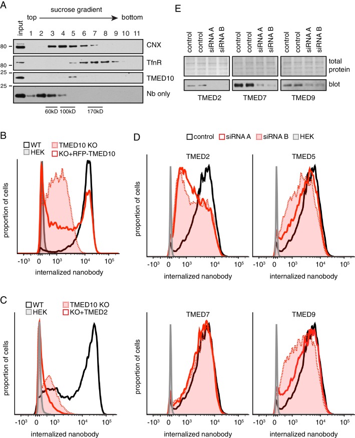
(A) Cell lysates prepared under non-denaturing conditions were separated by size on a 5–25% sucrose gradient, and analyzed by immunoblotting. Purified recombinant nanobody (18 kDa) peaks in fraction 2, as expected for a small monomeric protein. Despite a similar molecular weight, native TMED10 peaks in fraction five indicating its constitutive engagement in a higher molecular weight complex. For comparison, CNX (~90 kDa monomer) peaks in fraction 4, while the transferrin receptor (TfnR, dimer of two 85 kDa polypeptides) peaks in fractions 6–8. This suggests that native TMED10 is part of a ~ 100–125 kD complex. (B) Wild type (WT) cells, TMED10 knockout (KO) cells, KO cells transiently transfected with RFP-tagged TMED10, and HEK293 cells were assayed for their ability to internalize fluorescent nanobody for 1 hr in the presence of thapsigargin-induced ER stress. Nanobody fluorescence was measured by flow cytometry. RFP-tagged TMED10 is able to partially rescue nanobody internalization in ∆TMED10 cells. (C) GFP-PrP*-expressing cells lacking endogenous TMED10 (KO) were transiently transfected with myc-tagged TMED2 and assayed for their ability to internalize fluorescent nanobody for 2 hr in the presence of thapsigargin-induced ER stress. TMED2 cannot rescue the Nb-uptake deficiency of ∆TMED10 cells, and seems to even have a slight dominant-negative effect (i.e., further reduced Nb uptake). (D) Cells expressing GFP-PrP* were depleted of the indicated TMED family members using two independent siRNA oligonucleotides for 72 hr, and subsequently assayed for their ability to internalize fluorescent nanobody from the medium over a 100 min time period under non-stressed conditions. Although not shown here, no effect was seen for siRNA-treated TMED1, TMED3, TMED4, and TMED6. (E) Knockdown efficiencies for panel D were checked by immunoblotting for TMED2, TMED7, and TMED9. We were unable to obtain a suitable antibody for TMED5.