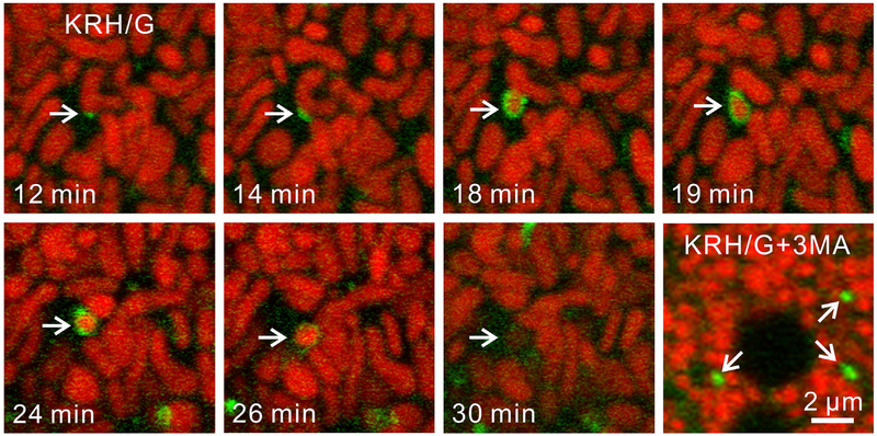
Fig. 2. Type 1 mitophagic sequestration of a polarized mitochondrion during nutrient deprivation.
GFP-LC3 transgenic hepatocytes were labeled with ΔΨ-indicating TMRM. Panels labeled 12 through 30 min represent a single experiment. After the incubation of hepatocytes in nutrient deficient Krebs-Ringer-HEPES medium containing 1 μM glucagon (KRH/G), a PAS associates with a TMRM-labeled mitochondrion (12 min) and grows into a phagophore that surrounds and sequesters a portion of the target organelle (arrows, 14–19 min). Note coordinate mitochondrial fission (18–19 min) and that mitochondrial depolarization (loss of TMRM fluorescence) does not occur until after sequestration (30 min). In a separate experiment under the same conditions but in the presence of the PI3K inhibitor 3-methyladenine (3 MA) that blocks type 1 mitophagy, PAS persist in apparent association with mitochondria (arrows, lower right panel). Adapted from references 9,13. Abbreviations: GFP-LC3, green fluorescent protein-microtubule-associated protein 1A/1B-light chain 3; ΔΨ, membrane potential; TMRM, tetramethylrhodamine methyester; PAS, preautophagic structure; PI3K, phosphoinositide-3-kinase.

