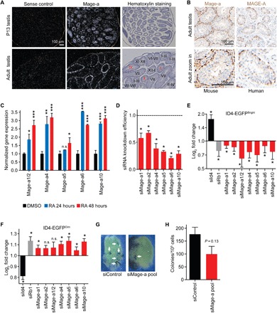
Fig. 2. Mage-a genes promote the maintenance of SSCs.

(A and B) In situ hybridization (A) and immunohistochemistry (B) show that Mage-a genes are expressed in early stages of spermatogenesis. Staining was performed using human and mouse anti-MAGE-A antibodies that recognize multiple MAGE-A proteins, including mouse Mage-a1/Mage-a2/Mage-a3/Mage-a5/Mage-a6/Mage-a8. (C) Primary spermatogonial stem cell cultures were treated with dimethyl sulfoxide (DMSO) or RA for 24 or 48 hours before expression of Mage-a genes were detected by RT-QPCR (n = 3). Data are means ± SEM. (D to F) Mage-a genes are required to maintain ID4-EGFPbright stem cells in primary SSC cultures. Knockdown efficiency after a 24-hour transfection is shown (D). Log2 fold change of ID4-EGFPbright (E) and ID4-EGFPdim (F) cells after knockdown of indicated genes (n = 3 biological replicates on a single ID4-EGFP SSC cell line). Data are means ± SD. (G and H) Mage-a genes are required for robust stem cell repopulation of testis (n = 3 technical replicates). Note that siMAGE-A depletes multiple Mage-a genes. Data shown are mean ± SEM. P values determined by Student’s t test, *P < 0.05, **P < 0.01, ***P <0.001. n.s., not significant.
