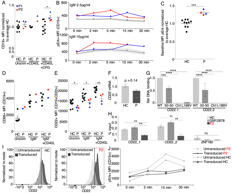
Fig. 2. Hyperresponsive phenotype of mutant IKAROS B cells is rescued by CD22 overexpression.
(A) CD19+ MFI on B cells from the CFSE assay in Fig. E6. (B) PBMCs stimulated with 2.5μg/ml or 10μg/ml anti-IgM-IgA-IgG were stained for pErk. Shaded area marks one standard deviation above and below the mean of the HC (2 repeats). (C) Baseline MFI of pErk in primary B cells for patients versus HC normalized to the average of HC. (D) PBMCs stimulated with anti-IgM-IgA-IgG 10μg/ml alone or or anti-IgM-IgA-IgG 10μg/ml + CD40L 1μg/ml for 24hrs were stained for CD69 (1 repeat) and (E) CD22 (2 repeat). (F) CD22 mRNA expression normalized to GAPDH and Actin for P and seven HC for 3 repeats. (G) Relative quantification of DNA binding on CD22 specific EMSA probes, normalized to WT binding (3 repeats). (H) ChIP-qPCR of IKAROS in EBV transformed B cells of P2, HC and GM12878 human B lymphocytes for CD22 and ZNF180 (not bound by IKAROS) (3 repeats). (I) CD22 MFI on CD19+ primary B cells after transduction with hCD22 for P2 and HC. (J) pErk MFI on hCD22 transduced CD19+ primary B cells after stimulation with 10μg/ml anti-IgM-IgA-IgG (1 repeat). Mean±SEM. **** = p<0.0001, *** = p<0.001 ** = p<0.01, * = p<0.05.

