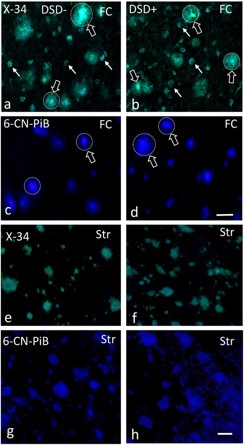
Figure 2.
Photomicrographs of frontal cortex X-34 (a, b) positive plaques (unfilled white arrows) and NFTs (filled white arrows) and 6-CN-PiB (c, d) positive plaques (unfilled white arrows) in a 44 year-old female non-demented (a, c) and a 59 year-old female demented (b, d) subject with DS. Note the intense fluorescence for both dyes (unfilled arrows) in the center of the plaques (dotted circles), characteristic of classic cored/neuritic plaques. Images showing X-34 (e, f) and 6-CN-PiB (g, h) positive plaques in the caudate of a 46 year-old male non-demented and a 59 year-old female demented DS case. Note the diffuse, less intense pattern of X-34 and 6-CN-PiB histofluorescence and the lack of a center core compared to FC plaques (a-d). Abbreviations: DSD−, DS without dementia, DSD+, DS with dementia, FC, frontal cortex, Str, striatum. Scale bars in d and h = 50 μm.

