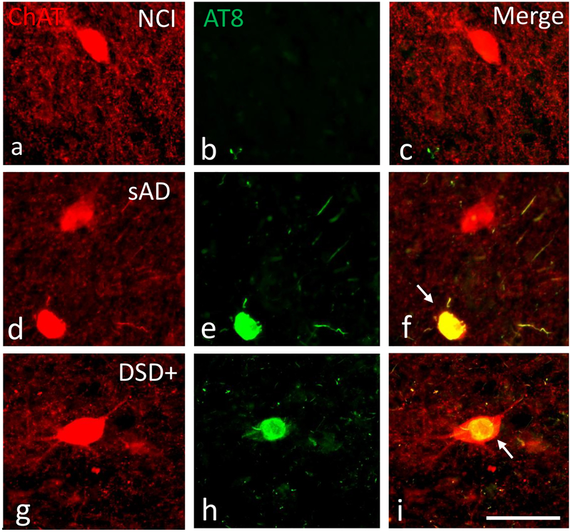
Figure 8.
Immunofluorescence showing striatal neurons single labeled for ChAT (red) and AT8 (green) and merged (yellow) images from a 71 year-old female non-cognitively impaired (a-c), a 76 year-old female severe AD (d-f) and a 46 year-old male demented cases with (g-i) case. Note that AT8 reactive within cholinergic neurons (yellow) in the AD and in demented case with DS, but not in the non-cognitively impaired aged subject. Of particular interest is the globose and shrunken appearance of the cholinergic tangle-bearing neuron (white arrow) in AD compared to the relative normal morphology despite NFT pathology within cholinergic perikarya in a demented subject with DS. Abbreviations: NCI, non-cognitive impairment, sAD, severe AD. The 50μm scale bar in j applies to all panels.

