
Fig. 4. BRUCE interacts with PRP19 which is required for FANCD2 mono-ubiquitination and foci formation.
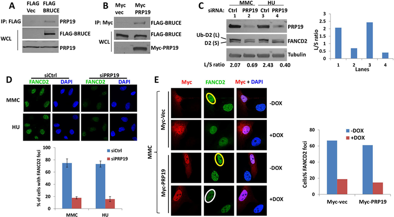
(A) U2OS cells stably expressing of FLAG-BRUCE were lysed and immunoprecipitation was carried out with anti-FLAG antibody. Elution was subjected to SDS-PAGE and immunoblotted for PRP19.
(B) U2OS cells that stably expressed FLAG-BRUCE were transfected with Myc-vector or Myc-PRP19 expression vector, and immunoprecipitation was carried out with anti-MYC antibody. The eluate was subjected to SDS-PAGE and immunoblotting against FLAG (BRUCE).
(C) U2OS cells were depleted of PRP19 by siRNA and treated with MMC or HU for 24 hrs with a final concentration at 1 μm and 2 mM respectively. Cells were collected and subject to immunoblot against BRUCE, FANCD2 and Tubulin, with the intensity of the ratio between FANCD2 large band (Ub-D2) and small band (D2) quantified with ImageJ software and shown to the right.
(D) U2OS cells were depleted of PRP19 by siRNA and treated with 1 μm MMC or 2 mM HU for 24 hours. Cells were fixed and stained for FANCD2 foci, representative images are present with the quantification of FANCD2 foci shown below. Bars represent SD from a triplicate experiment.
(E) U2OS cells were treated with DOX and transfected with MYC vector or MYC-PRP19 expression vector. After treatment with 1 μm MMC for 24 hours, cells were fixed and immunostained for MYC (Red) and FANCD2 (Green). Quantification of FANCD2 foci was shown to the right. Circle in yellow and white showing positive and negative nuclear FANCD2 foci, respectively.