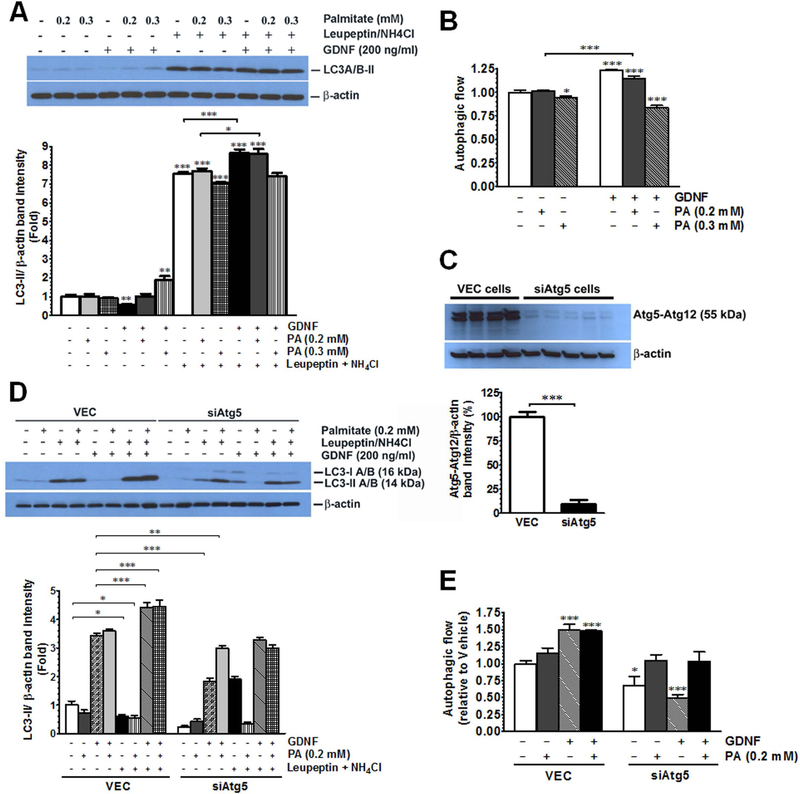
FIG. 2.
GDNF enhances autophagic flux in rat hepatocytes and primary mouse primary mouse hepatocytes in vitro (A) Western blot analysis of LC3 levels in VEC (empty vector-infected) and siAtg5 (Atg5 siRNA infected) RALA255–10G rat hepatocytes pre-cultured for 1 h in medium supplemented with or without the lysosomal inhibitors leupeptin and ammonium chloride followed by 4 h in medium supplemented with or without GDNF and palmitate (PA), and (B) plot of autophagic flow for GDNF and PA treated cells relative to cells treated with vehicle only. Plotted are means + SEM (***, P<0.001; *, P<0.05. n = 3). (C) Western blot comparing Atg5 protein expression levels between VEC and siAtg5 RALA255–10G rat hepatocytes. Plotted are means + SEM (***, P<0.001. n= 4–5). (D) Western blot analysis of LC3 levels in primary mouse hepatocytes pre-cultured for 1h in medium supplemented with or without the lysosomal inhibitors leupeptin and ammonium chloride followed by 4h in medium supplemented with or without GDNF and palmitate (PA), and (E) plot of autophagic flow for GDNF and PA treated cells relative to cells treated with vehicle only. Plotted are means + SEM (***, P<0.001; *, P<0.05. n = 3).

