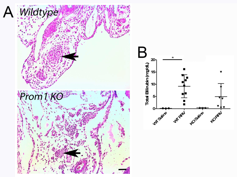
Figure 2.
Comparison of Prom1 WT and KO Cholestasis Two Weeks Following RRV Inoculation. (A) Hematoxylin and eosin staining of WT and KO extrahepatic structures (arrows denote obliterated bile duct. Scale bar = 25 µm. (B) Serum total bilirubin levels of WT and KO mice two weeks following saline or RRV injection. Abbreviations: KO, knockout; PV, portal vein; RRV, Rhesus rotavirus; WT, wildtype. * p<0.05.

