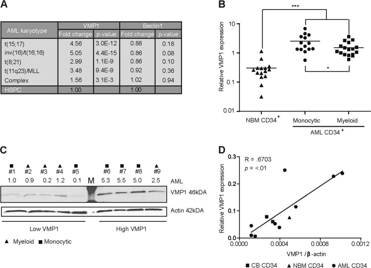
Fig. 1. VMP1 expression is increased in a subset of AMLs.
a Expression of VMP1 and Beclin-1 in mononuclear primary AML cells acquired from the publicly available expression dataset Bloodspot. HSPC is defined as the combined fractions of HSC, MMP, CMP, GMP and MEP. b Gene expression of VMP1 determined by quantitative RT-PCR in normal bone marrow (NBM) CD34+ cells (n = 15), AMLs with myeloid (n = 17) or monocytic characteristics (n = 14). c Western Blot showing variation in VMP1 protein levels in different AMLs (n = 9), β-actin was used as control and VMP1/β-actin levels are shown relative to AML1. Triangles, squares or circles indicate AMLs with a myeloid or monocytic, respectively. d Correlation between VMP1 mRNA levels and protein levels in primary AML CD34+ cells (n = 9, square symbols), CB CD34+ cells (n = 2, circles) and NBM CD34+ cells (n = 1, triangle). Error bars represent SD; * or *** represents p < .05 or p < .001, respectively

