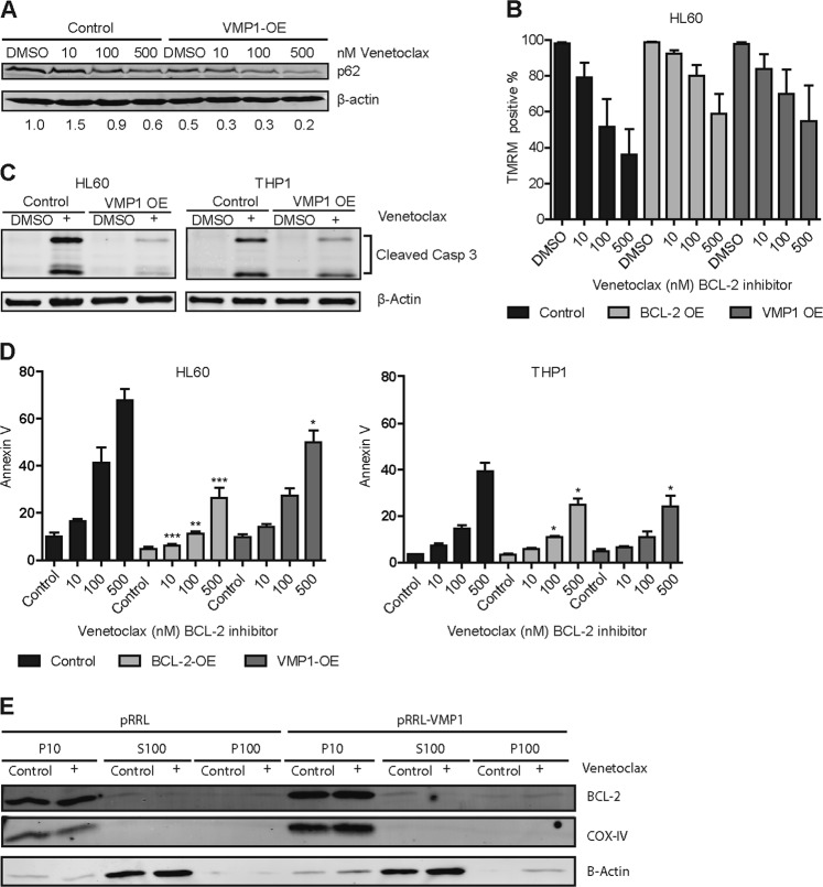
Fig. 6. Overexpression of VMP1 interferes with venetoclax induced apoptosis.
a Western blot of p62 in THP1 cells overexpressing VMP1 or control treated with different concentrations of venetoclax, β-actin was used as control. (n = 2) b Bar graph showing the percentage of TMRM positive cells. HL60 cells overexpressing VMP1, BCL-2 or control were incubated for 24 h with different concentrations of venetoclax. (n = 3) c Western Blot showing cleaved caspase 3 in HL60 and THP1 cells overexpressing VMP1 or control after 24 h incubation with 25 nM venetoclax (n = 2). d Leukemic cell lines with lentiviral overexpression of VMP1, BCL-2 or control were treated 24 h with venetoclax and apoptosis was measured by annexin-V staining with FACS (n = 4). e Western blot showing BCL-2, COX-IV, and β-Actin in the mitochondrial (P10), cytosolic (S100) or endoplasmic reticulum (P100) fractions of HL60 cells overexpressing VMP1 or control, treated with or without venetoclax. Error bars represent SD; *, ** or *** represents p < .05, p < .01 or p < .001, respectively

