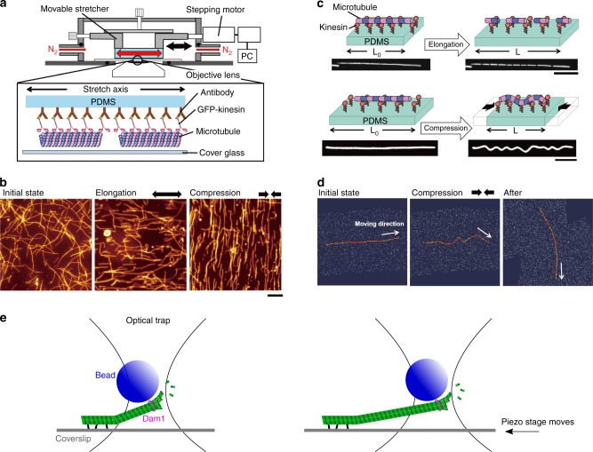
Fig. 3.
In vitro microtubules under mechanical stress. a Schematic diagram of an in vitro system to apply tension and compression to gliding microtubules on a kinesin-coated elastomer substrate (adapted from ref. 43). b Microtubules driven by surface immobilized kinesins align along maximal tension in vitro and conversely align against compression direction (adapted from ref. 43). c Fragmentation and buckling of microtubules at a stationary state induced by external tension and compression (adapted from ref. 90). e Using optical tweezer, growth of single microtubule is promoted when under tension along the direction of protofilaments (adapted from ref. 41, not to scale). d Microtubule aligns toward the direction that minimizes accumulated bending energy in silico (adapted from ref. 43)

