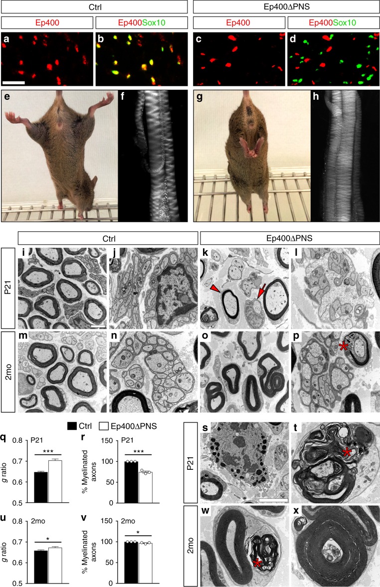
Fig. 1.
Peripheral neuropathy resulting from Ep400 deletion in Schwann cells (SCs). a–d Occurrence of Ep400 in SCs of sciatic nerves from control (a, b) and Ep400ΔPNS (c, d) mice at P21 as determined by co-immunofluorescence studies with antibodies against Ep400 (red) and Sox10 (green) to prove efficient SC-specific deletion. Sox10-negative cells in the nerve retained Ep400 and may represent endoneurial fibroblasts, pericytes, endothelial cells, or immune cells. Scale bar: 25 µm. e–h Hindlimb clasping phenotype (e, g) and sciatic nerve hypomyelination (f, h) in Ep400ΔPNS (g, h) as compared to control (e, f) mice at P21. i–p, s, t, w, x Representative electron microscopic pictures of sciatic nerve sections from control (i, j, m, n) and Ep400ΔPNS (k, l, o, p, s, t, w, x) mice at P21 (i–l, s, t) and 2 months (2 mo) (m–p, w, x) in overview (i–p) and at higher resolution (s, t, w, x). Magnifications depict an activated macrophage (s) and various myelin abnormalities (t, w, x). Arrow, unmyelinated axon; arrowhead, hypomyelinated axon; asterisk, myelin debris. Scale bars: 2.5 µm. q, r, u, v Determination of the mean g ratio (q, u) and the number of myelinated axons as percentage of total axons with a diameter ≥1 µm (r, v) in ultrathin sciatic nerve sections of control (black bars) and Ep400ΔPNS (white bars) mice at P21 (q, r) and 2 mo (u, v). All large caliber axons were myelinated in control mice. Statistical significance was determined by unpaired, two-tailed Student’s t test (*P ≤ 0.05; ***P ≤ 0.001). Exact values are listed in Supplementary Tables 1 and 2 and source data are provided as a Source Data file

