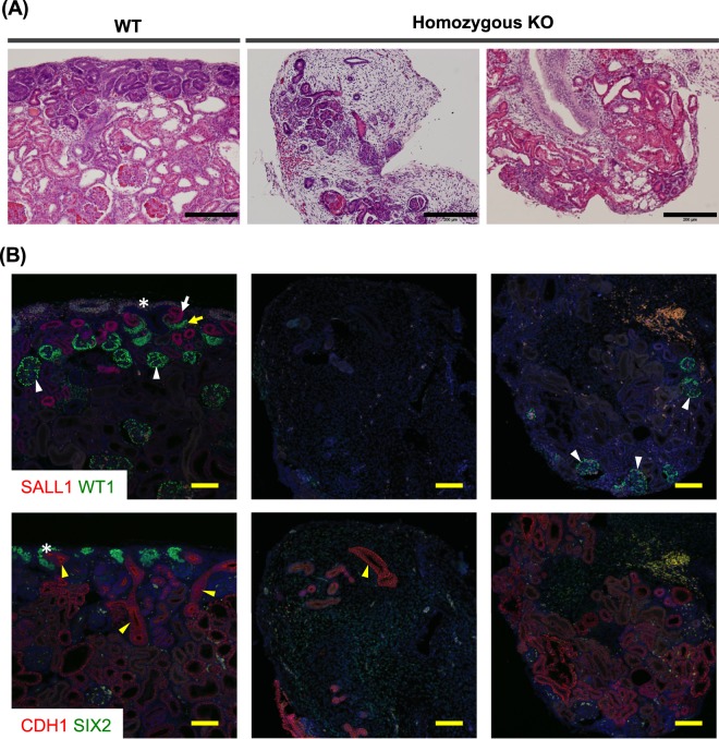
Figure 4.
Hypoplastic kidneys of F1 progeny carrying SALL1 mutations. Left panels: Kidney tissue of a WT foetus (W307-3). Middle and right panels: Kidney tissue of homozygous SALL1-KO F1 progeny (W307-8; 1 bp ins/13 bp del and W307-11; 1 bp ins/660 bp del) at day 40 of gestation. (A) Histological examination of foetal kidneys by haematoxylin-eosin staining. Homozygous KO foetuses showed kidneys that were disorganized and severely hypoplastic kidneys compared to those of WT foetuses. Black scale bars: 200 µm. (B) Immunofluorescence analysis of kidneys of homozygous SALL1-KO F1 progeny. Upper panels: SALL1, which was expressed in nephron progenitors (*) and distal nephrons (white arrow) in WT foetuses, was absent in homozygous KO foetuses (middle and right panels). WT1 was expressed in proximal nephrons (yellow arrow) and glomeruli (white arrowheads) in WT foetuses, whereas residual glomeruli (white arrowheads) were detected in KO foetuses. Lower panels: SIX2, which was expressed in nephron progenitors (*) in WT foetuses, was absent in homozygous KO foetuses. Ureteric buds (yellow arrowheads), which expressed CDH1 (E-cadherin), were detected only residually in KO foetuses. Yellow scale bars: 100 µm.

