Figure 2.
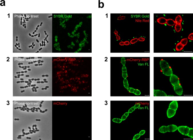
Fluorescence imagining of a phage and a phage antireceptor (RBP) binding to S. thermophilus. Strain STCH_12, phage CHPC951, a fluorescence derivative of the RBP of phage CHPC951, and a purified mCherry protein were used in the experiments. (a) Imagining under a conventional fluorescence microscope: (1) adsorption of SYBR Gold DNA-labelled phages (green) to the host strain; (2) adsorption of mCherry-tagged RBP (red) to the bacterial cells; (3) no adsorption of mCherry proteins to the bacterial cells. (b) Super-resolution structured illumination microscopy (SIM) imagining: (1) bacterial cells were labelled with Nile Red (red) and mixed with SYBR Gold-DNA labelled phages (green); (2) bacterial cells were labelled with Van FL (green) and mixed with mCherry-tagged RBP (red); (3) bacterial cells were labelled with Van FL (green) and mixed with mCherry proteins (red). Scale bars: 1 µm.

