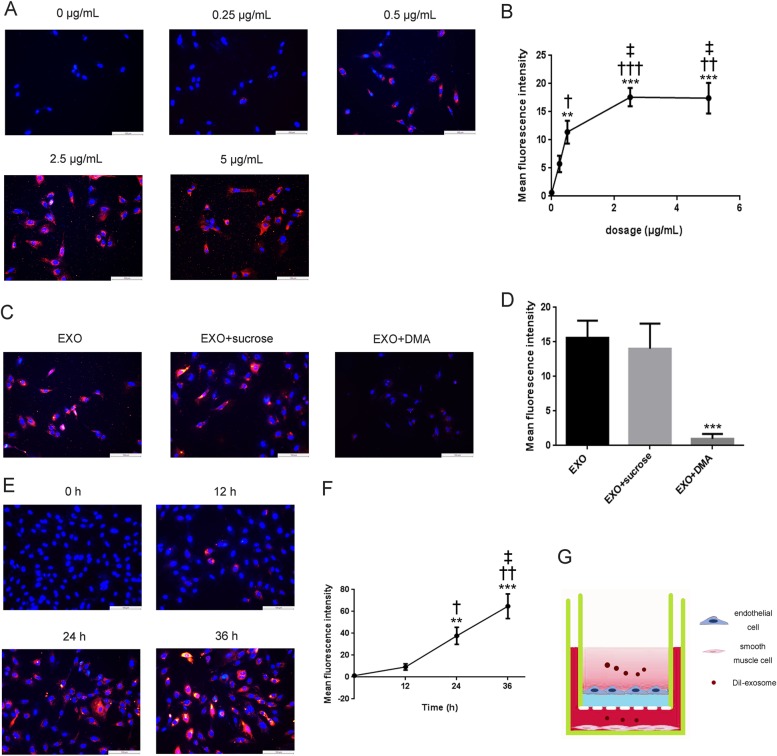
Fig. 2. Cellular uptake and endocytic mechanism in HA-VSMC and permeation of T1-EXO across endothelial cell layer.
a, b Representative fluorescent microscopy of HA-VSMCs that were exposed to DiI-labeled exosomes derived from M1M for 6 h. *P < 0.05, versus 0 μg/mL group; †P < 0.05, versus 0.25 μg/mL group; ‡P < 0.05, versus 0.5 μg/mL group. n = 3, each group. c, d Effect of sucrose and DMA on cellular uptake of DiI-labeled T1-EXO in HA-VSMCs. Fluorescence intensity of HA-VSMCs exposed to 2.5 μg exosomes/mL without pre-incubation with any inhibitors representing the maximum internalized amount of DiI-labeled T1-EXO served as a control. *P < 0.05, versus EXO group. n = 3, each group. e, f Representative fluorescent images of HA-VSMC after treatment with DiI-labeled T1-EXO. *P < 0.05, versus 0 h group; †P < 0.05, versus 12 h group; ‡P < 0.05, versus 24 h group. n = 3, each group. g Schematic diagram of permeation of T1-EXO across endothelial cell monolayer into smooth muscle cells. All data were expressed as mean ± SEM from three individual experiments

