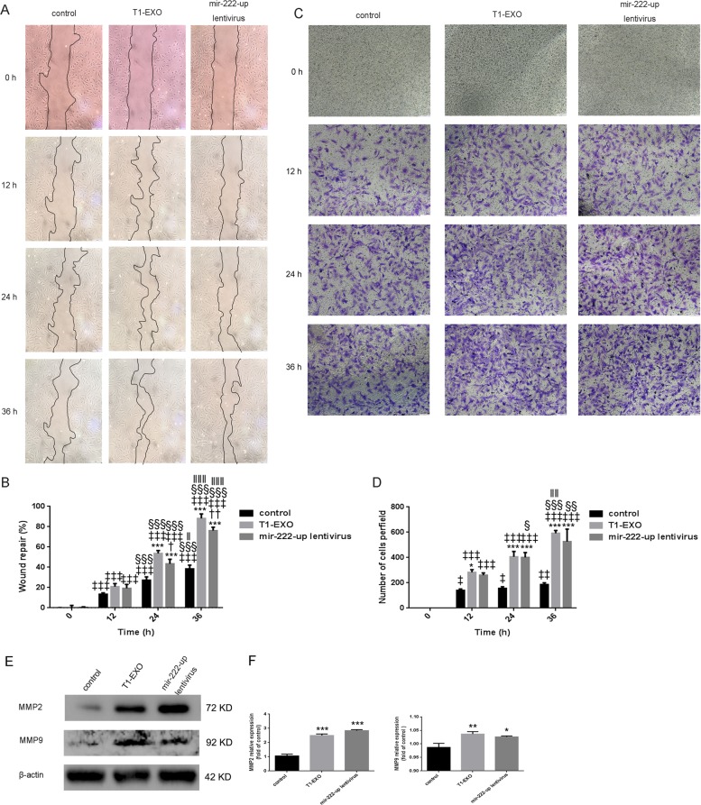
Fig. 5. T1-EXO and miR-222 enhanced HA-VSMC migration.
a Quiescent scratched HA-VSMCs were treated or untreated with T1-EXO (25 μg/mL) in FBS-free medium for 36 h. VSMCs infected with mir-222-up lentivirus served as a positive control. Phase-contrast microscopy images showing the migratory capacity of VSMCs under different treatment. b Diagram showed quantification of cell migration rate over time. *P < 0.05, versus control group; †P < 0.05, versus T1-EXO group; ‡P < 0.05, versus 0 h group; §P < 0.05, versus 12 h; ‖P < 0.05, versus 24 h group. n = 3, each group. c A transwell assay was performed to assess HA-VSMCs migration. d Diagram showed quantification of migrated cells through transwell chamber. *P < 0.05, versus control group; ‡P < 0.05, versus 0 h group; §P < 0.05, versus 12 h; ‖P < 0.05, versus 24 h group. n = 3, each group. e, f T1-EXO and miR-222 improved the expression of proteins involved in smooth muscle cell migration: MMP2 and MMP9. β-actin expressions served as internal control. The control group was set to 1. *P < 0.05, versus control group. n = 3, each group. All data were expressed as mean ± SEM from three individual experiments

