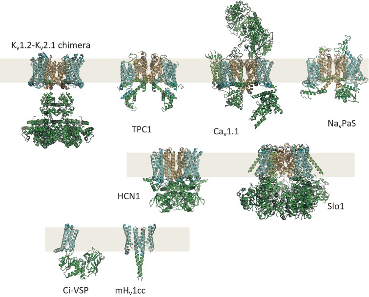
Figure 2.
Gallery of protein structures of VGICs and VSP (taken from ref. 157). Kv1.2–Kv2.1 chimera: human voltage-gated potassium channel (PDB ID: 2R9R), TPC1: plant two-pore cation channel (PDB ID: 5E1J), Cav1.1: human skeletal muscle-type voltage-gated calcium channel (PDB ID: 5GJV), NavPaS: insect voltage-gated sodium channel (PDB ID: 5X0M), HCN1: human hyperpolarization-activated cyclic nucleotide-gated channel (PDB ID: 5U6O), Slo1: large conductance calcium-activated voltage-gated potassium channel from Aplysia (PDB ID: 5TJ6), Ci-VSP: sea squirt voltage-sensing phosphatase (full length model based on coordinates from PDB ID: 3AWF and PDB ID: 4G7V), mHv1cc: mouse voltage-gated proton channel in a chimeric form containing part of S2–S3 from Ci-VSP and the coiled coil structure of GCN4 (dimer model based on coordinates from PDB ID: 3WKV). NavPaS, Kv1.2–Kv2.1 chimera and Slo1 have four VSDs within a tetramer. TPC1 has four VSDs within a dimer. Ci-VSP has a single VSD. mHv1cc has two VSDs within a dimer.

