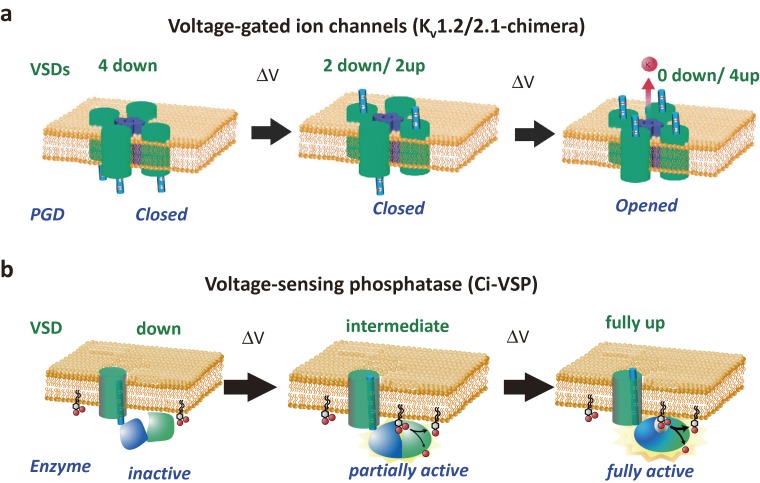
Figure 6.
Operation of voltage-sensor containing proteins. In most of voltage-gated ion channels, multiple voltage sensor domains (green) need to take up-state (activate state) until the pore is opened (upper pictures (a)). This is one of the reasons why the voltage dependence of conductance increase in voltage-gated ion channels is very sharp, as represented by voltage-dependent conductance of nerve membranes as first demonstrated by Hodgkin and Huxley. However, it should be noted that some VGICs require only one or two VSDs in the activated state to have the PGD in an open state. Lower pictures (b) show a current model of operation mechanisms of VSP. In contrast with four homologous repeats or subunits in VGICs, single VSD (green) is connected with the cytoplasmic region, which consists of the phosphatase domain (PD)(blue) and C2 domain (light green) in VSP. Upon partial activation of VSD, some phosphatase activity emerges, whereas the enzyme is fully activated upon full activation of VSD. PD has a flexible structure and its association with the membrane is critical for full activation of the enzyme when the VSD is fully activated. This second step requires a special motif, called a “hydrophobic spine” (not drawn in the diagram).60) In both cases, illustration of the motion of S4 follows the classical idea of translation motion for the simplicity of the figure, rather than ideas of transporter model and helical screw model.158)

