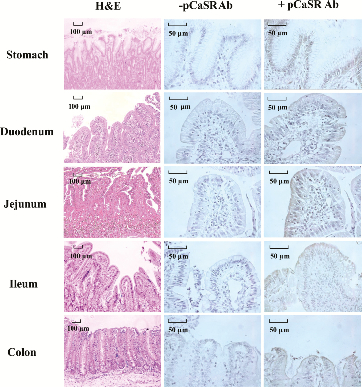
Figure 7.
Haemotoxylin and Eosin (H&E) staining and immunohistochemical staining of pCaSR protein in the stomach, duodenum, jejunum, ileum, and colon in the weaned piglets. Positive staining for pCaSR was indicated by brown deposits. −pCaSR represented negative control which the primary antibody was omitted from the staining procedure. Scale bar (black) for H&E staining represents 100 μm, and for immunohistochemical staining is 50 μm.

