Fig. 1.
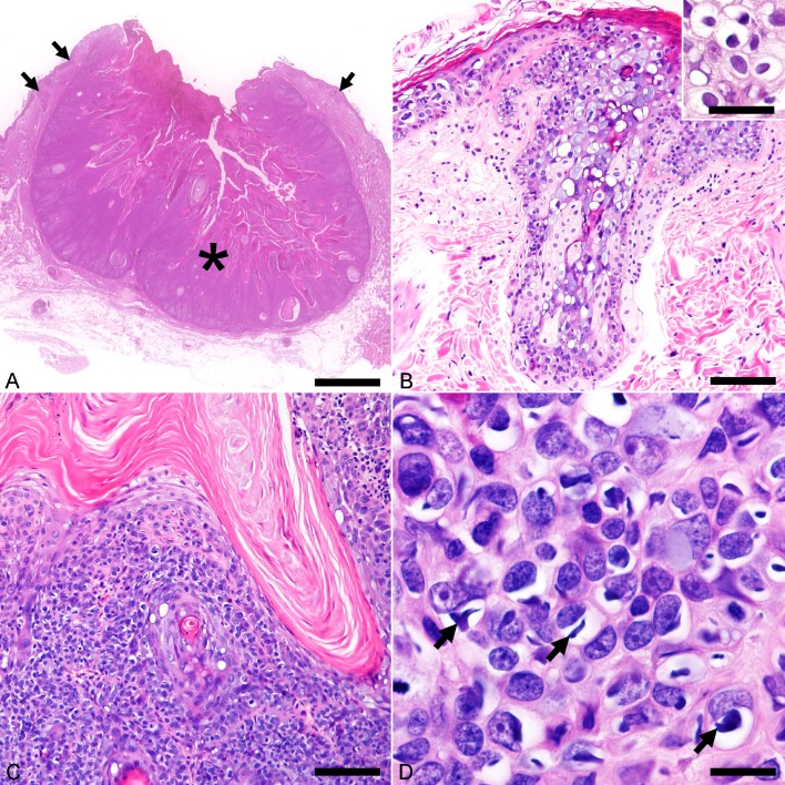
Histopathological features of Felis catus papillomavirus type 5 (FcaPV-5)-associated viral plaque and Bowenoid in situ carcinoma (BISC). Hematoxylin and eosin. (A) The neck mass consists of multicentric viral plaques (arrows) and a well-demarcated, keratinizing tumor lobule (asterisk). Bar, 2.5 mm. (B) The viral plaque consists of prominent epidermal and infundibular acanthosis accompanied by mild to moderate sebaceous hyperplasia. The affected suprabasal cells are swollen and have a blue-gray vacuolated cytoplasm. Bar, 100 µm. Inset shows koilocytes located in the stratum spinosum of acanthotic epidermis. Bar, 40 µm. (C) Cells of BISC proliferate in nests that are separated by thin fibrous stroma. Bar, 100 µm. (D) A high magnification shows cells of BISC display marked anisocytosis and anisokaryosis. Numerous cells have a clear swollen cytoplasm with spindle- to irregular-shaped, basophilic cytoplasmic bodies (arrows). Bar, 15 µm.

