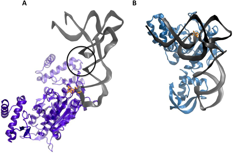
Figure 7: Substrate tRNA-binding modes of TrmD and Trm10.

(A) Co-crystal structure of TrmD with tRNA illustrates the binding of tRNA at the dimer interface. The two monomers forming the dimer are indicated in light and dark purple. Residues from the CTD of one monomer and the NTD from the second monomer interact with the core of the tRNA (indicated by the circle) and contribute to positioning the substrate G37 (orange) in the anticodon loop at the active site (PDB: 4YVI) 32,46 (B) Computational docking model of SaTrm10 with E. coli initiator tRNA predicts that the shape of full-length enzyme recognizes the L-shape of the tRNA which may help specifically identify the substrate core nucleotide R9 (orange)24.
