
Fig 4.
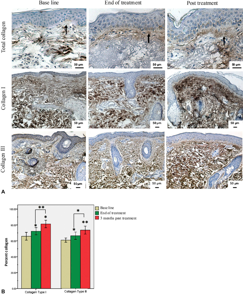
Increase in dermal collagen content in response to radiofrequency. A, Immunohisto-chemical staining of skin tissues at baseline (left), end of treatment (middle), and after RF treatment (right) for total collagen (top) and collagen types I (middle) and III (bottom). Increase in collagen band thickness at dermoepidermal junction was observed after RF treatment compared with baseline (arrows). B, Collagen level was measured and values presented as percentage of dermis-positive collagen. Data showed statistically significant increase in both collagen I and III in response to RF. *P ≤ .05; **P ≤ .001.