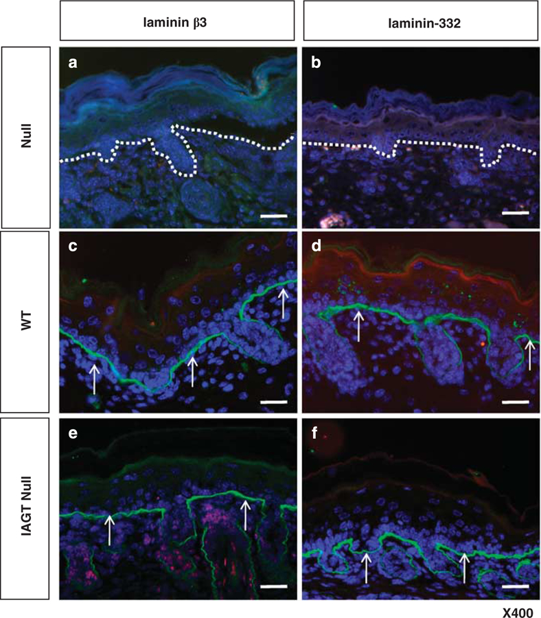
Figure 1.

Immunofluorescence analysis of skin samples from untreated homozygous LAMB3IAP mice (defined as ‘null’) (a, b), WT mice (defined as ‘WT’) (c, d) and homozygous LAMB3IAP mice treated by E8 IAGT with lentiviral-LAMB3 (defined as ‘IAGT null’) (e, f). No expression of either laminin-β3 or laminin-332 is seen in the BMZ in untreated null mice. The white dotted lines indicate the position of the basal lamina. (a, b) WT mice are positive for laminin-β3 and laminin-332 staining in a linear pattern along the basement membrane. (c, d) Treated null mice stain positive for laminin-β3 and laminin-332 at the basement membrane in a similar pattern to WT mice arrows (arrows, e, f). The sections were counterstained with 4’,6-diamidino-2-phenylindole to visualize the cells (blue) (scale bar=100 µm).
