
Figure 3.
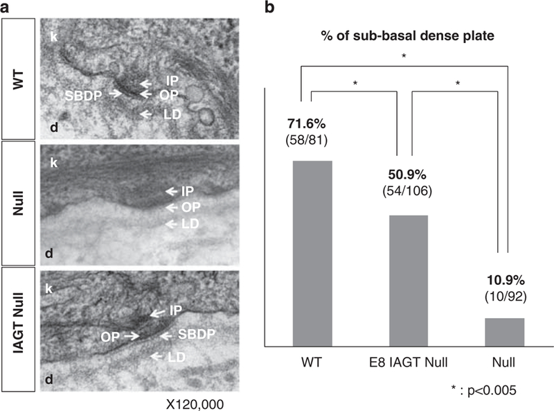
Transmission electron microscopy of dermo-epidermal junction. (a) Electron micrographs of skin sections demonstrate the junction between the basal keratinocytes (k) of the epidermis and the upper reticular dermis (d). In the WT mice, the hemidesmosomes are visible at the basal surface of the keratinocytes and are comprised of an inner plaque (IP), outer plaque (OP) and SBDP. The BMZ, consisting of the electron-opaque lamina lucida and the electron-dense lamina densa, runs parallel to the keratinocyte basal membrane. In homozygous LAMB3IAP mice (null), the hemidesmosomes are present but appear less distinct and the inner and OPs are more diffuse than in the WT mice, and the SBDP is absent. In homozygous LAMB3IAP mice treated by E8 IAGT with lentiviral-LAMB3 (IAGT null), the normal structures of the desmosomes are restored. (b) Bar graph demonstrating the percentage of hemidesmosomes (HD) with a demonstrable SBDP. A blinded examiner with expertise in skin electron microscopy counted the number of the HD and determined the presence or absence of the SBDP. *P-values o0.005 for positive SBDP comparison between WT, E8 IAGT null and non-treated null pups by using the Fisher’s exact test.