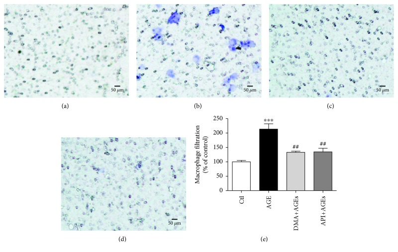
Figure 4.
DMA protects AGE-induced macrophage infiltration. RAW 264.7 macrophages at the upper compartment of the Boyden chamber were incubated with medium (a), 500 μg/mL AGEs (b), 10 μM DMA + 500 μg/mL AGEs (c), 10 μM API + 500 μg/mL AGEs (d) for 24 hrs. Filtered macrophages in the microporous membrane were stained by 0.1% crystal violet, and representative photos were taken under a microscope. (e) Macrophage counting. The stain was washed off by 30% acetic acid and measured at a wavelength of 580 nm. ∗∗∗ p < 0.001 vs. control; ## p < 0.01 vs. AGE treatment.

