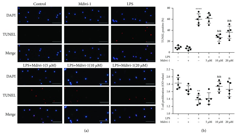
Figure 3.
Mdivi-1 protects CD4+ T cells against LPS-induced apoptosis. Isolated CD4+ T cells were activated by ConA (5 μg/mL), and then, Mdivi-1 (0, 10, and 20 μM) was administered 30 min prior to the LPS stimulation. (a) Representative photomicrographs of CD4+ T cells (around 20 cells) after colabeling TUNEL/DAPI and around 400 cells in each group evaluated to determine the percentage of TUNEL-positive cells. (b) Viability analysis of CD4+ T cells. Scale bars, 50 μm. The results are shown as the mean ± SD; n = 5. ∗∗ P < 0.01 vs. the control group (∗∗∗∗ P < 0.0001); & P < 0.05 vs. the LPS group (&& P < 0.01, &&& P < 0.001).

