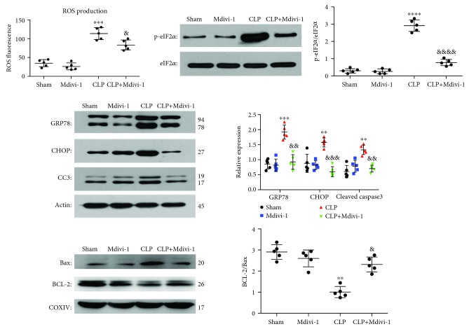
Figure 6.
Mdivi-1 reduced ROS production and prevented the induction of ER stress in CD4+ T cells after CLP. Mice were sacrificed at 24 h after CLP with or without Mdivi-1 treatment. (a) Quantification of ROS fluorescence intensity. (b) Western blot assay and quantitative analysis of p-eIF2α and eIF2α expression. (c) Western blot assay and quantitative analysis of GRP78, CHOP, and cleaved caspase-3 expression. (d) Western blot assay and quantitative analysis of BCL-2 and BAX expression. The results are shown as the mean ± SD; n = 5. ∗ P < 0.05 vs. the sham group (∗∗ P < 0.01, ∗∗∗ P < 0.001, and ∗∗∗∗ P < 0.0001); & P < 0.05 vs. the CLP group (&& P < 0.01, &&& P < 0.001, and &&&& P < 0.0001).

