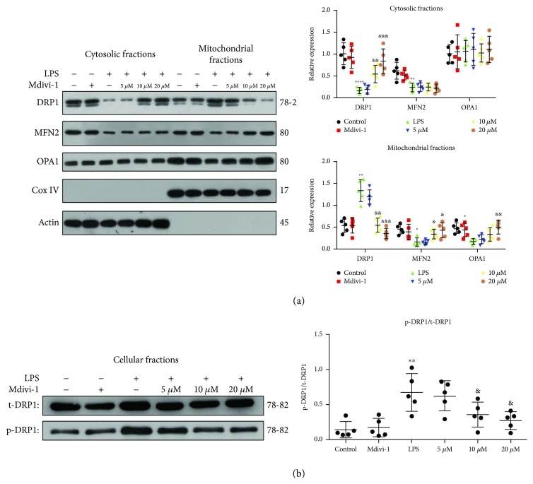
Figure 7.
Mdivi-1 reduced Drp1 translocation and Drp1 phosphorylation after the LPS treatment. Mdivi-1 (0, 10, and 20 μM) was administrated to activated CD4+ T cells 30 min prior to the LPS stimulation. (a) Western blot assay and quantitative analysis of mitochondrial MFN2, OPA1, and Drp1 expression and cytosolic MFN2, OPA1, and Drp1 expression. (b) Western blot assay and quantitative analysis of Drp1 phosphorylation. The results are shown as the mean ± SD; n = 5. ∗ P < 0.05 vs. the control group (∗∗ P < 0.01, ∗∗∗ P < 0.001, and ∗∗∗∗ P < 0.0001); & P < 0.05 vs. the LPS group (&& P < 0.01, &&& P < 0.001).

