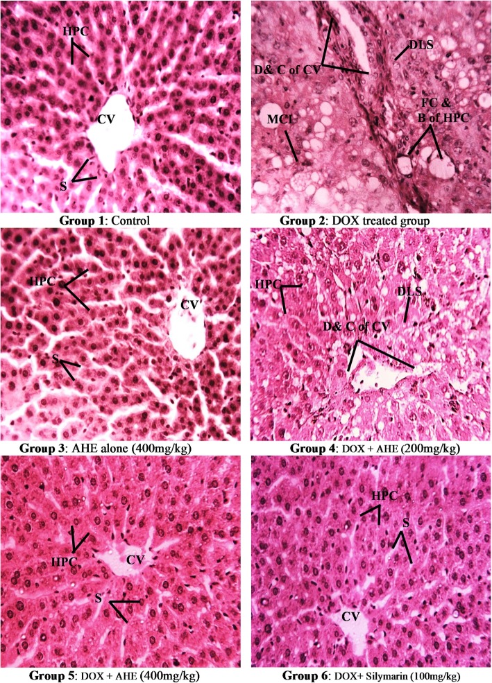
Fig. 3.
Histopathological examination of rat liver (H&E staining magnification 40X). Group 1: Liver section from control rats showing normal liver morphology. Group 2: DOX-treated rat liver section showing enhanced cellular lesions, loss of hepatic tissue structure arrangement and collection of inflammatory cells. Group 3: Represents liver section from AHE alone treated rats. Group 4: AHE low dose treatment results in minimal degenerations were observed. Group 5: AHE high dose treatment results in significant protection against DOX induced hepatic injury. Group 6: Showed protective effect of Silymarin treatment. AHE-A. hydaspica ethyl acetate fraction, DOX-Doxorubicin, HPC-Hepatocytes, CV-Central venule, MCI-Monocytes nuclear cells infiltrations, D&C-Dilation and congestion, FC & B- Fatty changes and ballooning, DLS- Degeneration of lobular shape, S-Sinosides, N-necrosis

