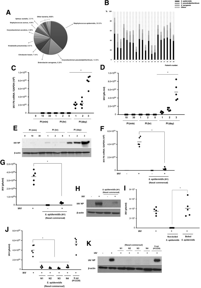
Fig. 1.
S. epidermidis exerts an antiviral effect against influenza A virus (IAV) infection in normal human nasal epithelial (NHNE) cells. a Bacterial species cultured from mucus samples obtained from the middle turbinates of healthy human subjects (n = 20) were identified via 16S rRNA gene sequencing. The distribution of the identified bacterial species is presented in the graph. b The relative abundance of nasal commensal, such as S. epidermidis, C. pseudodiphtheriticum, and E. aerogenes, in each subject. Bar graph presents the relative abundance of nasal commensal at the species level. NHNE cells from five healthy volunteers were inoculated with IAV at a multiplicity of infection (MOI) of 1. c IAV PA mRNA levels, normalized to cellular GAPDH transcript levels, were monitored by real-time PCR over the course of 3 days. d Viral titer of the infected NHNE cells was monitored using plaque assays. Results are presented as the mean ± standard deviation (SD) from five independent experiments. *p < 0.05 compared with mock-infected cells. e Levels of IAV nucleoprotein (NP) were monitored in NHNE cells following IAV infection using western blot analysis. Representative results are shown. PI post-infection. NHNE cells were inoculated with S. epidermidis N1 (MOI = 0.25) 8 h before IAV infection (MOI = 1). Subsequently, IAV PA mRNA levels (f), viral titers (g), and IAV NP protein levels (h) were assessed at 1 day post-infection (dpi) using real-time PCR, plaque assays, and western blotting, respectively. The mean viral titer was also measured in NHNE cells which were inoculated with boiled S. epidermidis and non-boiled S. epidermidis 8 h before IAV infection (i). Both mean viral titers (j) and IAV NP level (k) were determined at 1 dpi for NHNE cells infected with nasal mucus-derived S. epidermidis (N1, N2, N3, N4) and or non-pathogenic laboratory S. epidermidis ATCC 12228 prior to IAV infection. Real-time PCR and plaque assay results are presented as mean ± SD from five independent experiments. *p < 0.05 compared with the values for cells infected with IAV alone

