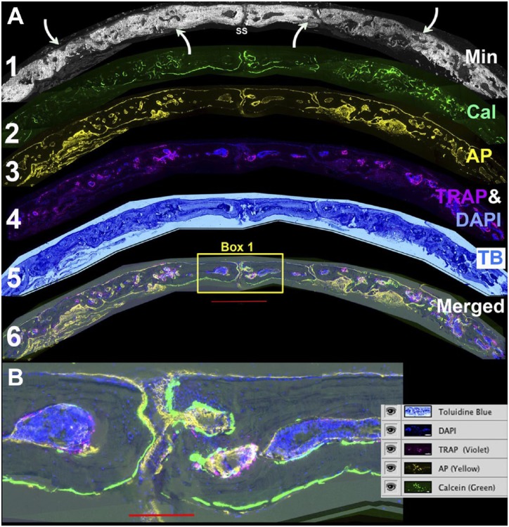
Figure 2.
Defining the osteogenic landscape of the repair field. (A) Individual layers of the stack are assembled to characterize the major features of the repair process. Layer 1: Accumulated mineral from which the overall orientation of the defect field is related to the sagittal suture (SS) and the boundaries of the repair are identified by the relative intensity of newly formed versus pre-existing mineralized bone (curved arrows). Layer 2: Calcein labeling of actively mineralizing bone surfaces. Both the accumulated mineral and mineral labeling signals are removed in the TRAP step. Layer 3: AP enzymatic activity. This step assesses the osteogenic activity of the repair process and is performed after the TRAP step. Layer 4. TRAP (violet) and DAPI (blue). The two are paired to emphasize the association of the endocortical bone surfaces with the bone marrow islands. The DAPI is also useful to identify cells with the bone matrix. Layer 5: TB. This stain provides a familiar chromogenic context for the fluorescent signals. Layer 6: Merging of all the cellular layers to show their relationship with active bone matrix forming surfaces. (B) Enlarged view of box 1 with a screen shot of the layer file. The green calcein label highlights the active periosteal and endosteal bone surfaces in the region of the SS. Most of the surfaces have an overlying AP (yellow signal). The bone marrow islands are intensely DAPI positive with TRAP-positive foci on the endocortical surface. The scale bar for Fig. 2A = 1000 µm and for Fig. 2B = 200 µm. Abbreviations: TRAP, tartrate resistant acid phosphatase; DAPI, 4′,6-diamidino-2-phenylindole; AP, alkaline phosphatase; TB, toluidine blue.

