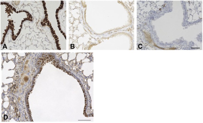
Figure 2.
Anti-mouse SCGB1A1 antibody specificity and airway β-tubulin expression. Immunohistochemistry was carried out using lungs of wild-type (A) and Scgb1a1(–/–) mice (kindly provided by Dr. Anil Mukherjee, NICHD) (B), and wild-type without primary antibody (C). Airways shown are large airways. Counterstained with hematoxylin. Scale bar: 50 µm for all panels. (D) Abundance of ciliated cells in mouse lung airway using C57BL/6N mouse. The airway shown is a large airway. Counterstained with hematoxylin. Scale bar: 50 µm.

