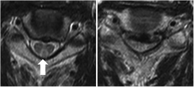
Figure 2.

Axial T2-wieghted MRI of a patients with degenerative cervical myelopathy and concomitant B12 deficiency. On the left, a characteristic reverse V-shaped hyperintensity is visible in the posterior column (arrow). On the right significant spinal cord compression is demonstrated. Reprinted with permission from Elsevier: The Spine Journal (Miyazaki et al8).
