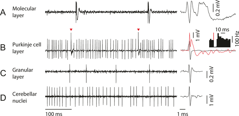
Fig. 6.
Neural activity patterns in each layer of cerebellar cortex and deep cerebellar nuclei. (a) In molecular layer, complex spike from Purkinje cell dendrite can be isolated. Zoomed-in view of complex spike waveform (right) shows long tail with spikelets, which can be detected as a low-frequency sound in audio monitor. (b) In Purkinje cell layer, simple spike and complex spike (red arrowhead and red trace) can be simultaneously isolated. Simple spike firing (y-axis of the inset histogram is firing rate) pauses for 10–30 ms after each complex spike (red line). (c) In granular layer, a low firing rate cell (putative Golgi cell) can be isolated. Complex spikes should not be detectable in this layer. (d) In deep cerebellar nuclei, cells with high spontaneous firing rate are often isolated (putative excitatory projection neuron). The cerebellar nuclei should be separated from the cerebellar cortex by white matter (which lacks prominent spiking activity and sounds quiet on the audio monitor)

