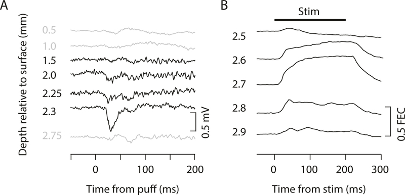
Fig. 7.
Functional mapping of cerebellar cortex and nuclei. (a) LFP recordings at different depths through cerebellar cortex during periocular stimulation (air puff). Region with periocular climbing fiber receptive fields is indicated by large negative deflection in LFP signal following air puff to eye. (b) Eyelid traces during microstimulation at different depths through DCN. The eyeblink region of DCN is indicated by the location at which microstimulation at currents less than approximately 10 μA produces large and reliable eyelid closure that is sustained for the duration of the stimulation (indicated by black line at top). Used with permission from [12]

