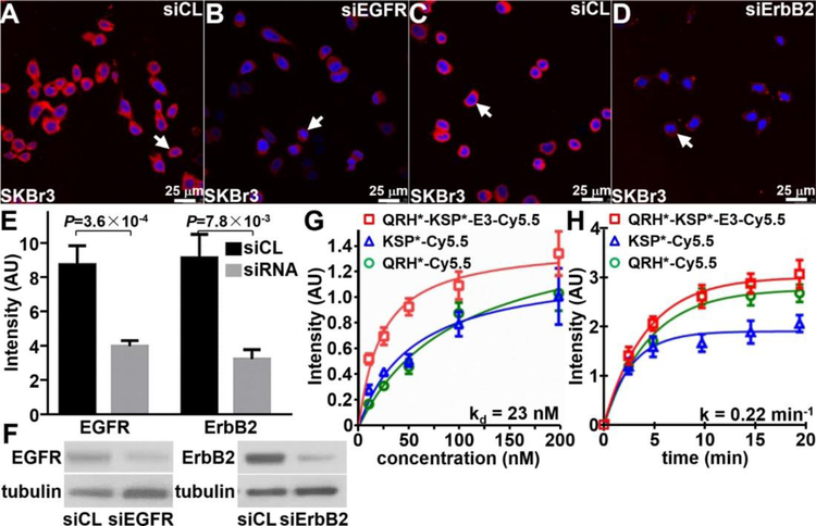
Figure 3. Characterization of peptide heterodimer.
In siRNA knockdown experiments, QRH*-KSP*-E3-Cy5.5 (red) shows significantly greater binding to the surface (arrows) of A) siCL (control) SkBr3 cells compared with that for B) siEGFR (knockdown) cells. Similar results were found for C) siCL and D) siErbB2 (knockdown) cells. E) Quantified results show significantly greater intensity for siCL versus siEGFR and siCL versus siErbB2, P=3.6×10⁻⁴ and P=7.8×10⁻³, respectively, by unpaired t-test. The mean value was calculated from 5 cells chosen randomly from 3 images collected independently. F) Western blot shows EGFR and ErbB2 expression in control and knockdown cells. G) The apparent dissociation constant (binding affinity) for QRH*-KSP*-E3-Cy5.5 was found to be kd = 23 versus 98 and 54 nM for QRH*-Cy5.5 and KSP*-Cy5.5. H) The apparent association time constant for QRH*-KSP*-E3-Cy5.5 was found to be k = 0.22 min−1 (4.5 min) versus 0.21 min−1 (4.8 min) and 0.35 min−1 (2.9 min) for QRH*-Cy5.5 and KSP*-Cy5.5. Results for each measurement are representative of 3 independent experiments.

