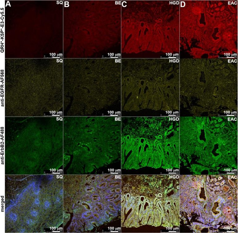
Figure 7. Specific binding of peptide heterodimer to Barrett’s neoplasia.
On representative confocal microscopy images of human esophageal specimens ex vivo, QRH*-KSP*-E3-Cy5.5 (red) shows minimal staining to A) squamous (SQ) and B) Barrett’s esophagus (BE), and increased intensity with C) high-grade dysplasia (HGD) and D) esophageal adenocarcinoma (EAC). Similar results were found with AF568-labeled anti-EGFR antibody (yellow) and for AF488-labeled anti-ErbB2 antibody (green). Merged images show co-localization of peptide and antibody binding.

