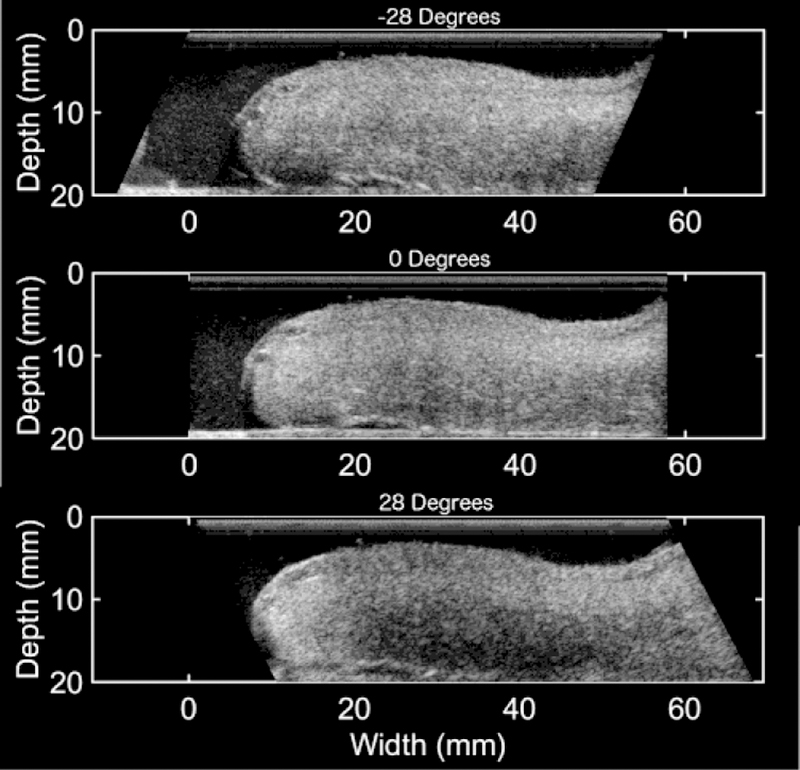
Figure 4.

B-mode images at −28, 0, and 28 degrees with respect to the transducer face of an ex vivo anterior cervix (proximal end on the right and distal end on the left). The brightness of the image is visibly different for each angle. The decreasing brightness from −28 degrees to 28 degrees corresponds to an increase in BSPD.
