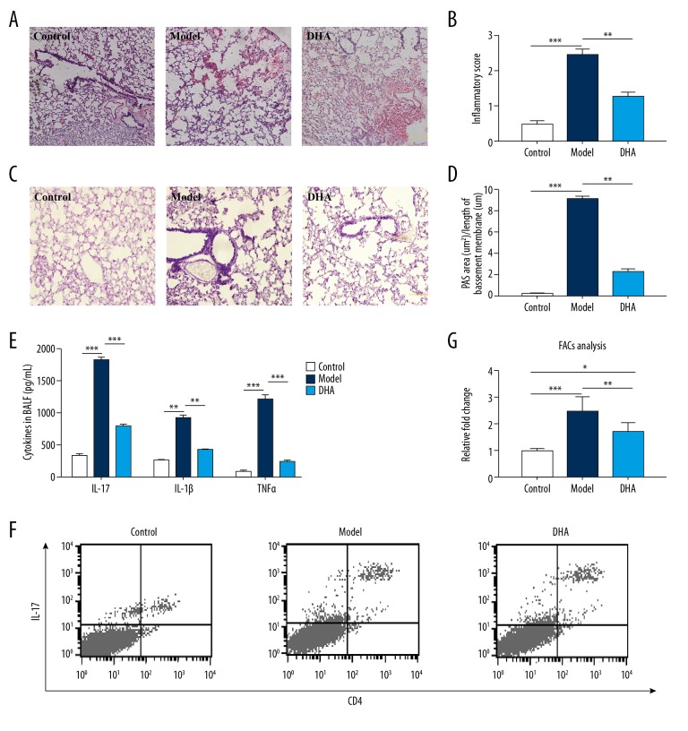
Figure 2.
DHA treatment ameliorates lung function and inflammation by lowering Th17 cytokine secretion and cells in BALF. (A) Micrographs of H&E staining of lung tissue sections (200× magnification, scale bar=50 μm, n=5). (B) Statistics for inflammation score of 2A. (C) Micrographs of PAS staining of lung tissue sections (200× magnification, scale bar=50 μm, n=5). (D) Statistics for degree of mucus accumulation [PAS+ area (μm 2) divided by the length of basement membrane (μm)] of 2C. (E) Inflammatory cytokine (IL-17, IL-10, IL-1β, and TNFα) changes in BALF in the Control, Model, and DHA group mice by ELISA (n=7). (F) Representative density plots of CD4+IL-17+ cells in BALF are shown (n=5). (G) Relative ratio of CD4+IL-17+ cells in 2D.* P<0.05, ** P<0.01, *** P<0.001. BALF – bronchoalveolar lavage fluid; H&E – hemotoxin and eosin; PAS – periodic acid-Schiff; DHA – dihydroartemisinin; OVA – ovalbumin.

