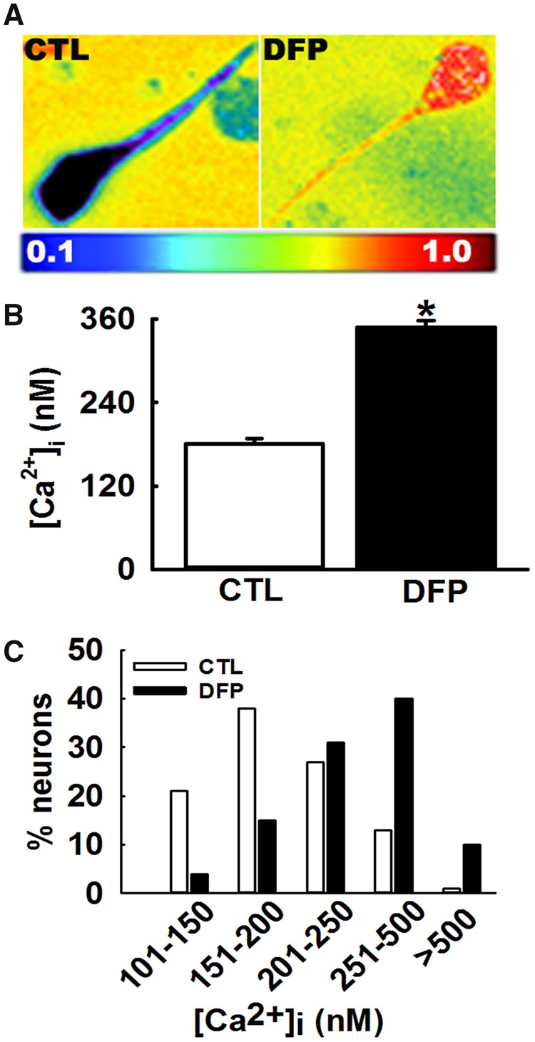
Figure 2.

Protracted hippocampal [Ca2+]i following repeated, low-dose OP-DFP exposure in rats. A, Pseudocolor ratiometric images of representative acutely isolated hippocampal CA1 neurons from age-matched control and DFP rat. Control neurons had bluish color that corresponds to lower Fura-2 ratio. DFP neurons had orange-red color that corresponds to higher Fura-2 ratio. B, Bar graph comparing [Ca2+]i in control versus DFP neurons. CA1 hippocampal neurons acutely isolated at 3 months following repeated, low-dose DFP exposure exhibited an increased Fura-2AM 340/380 ratios indicative of [Ca2+]i that were significantly higher compared with the calibrated [Ca2+]i from age-matched control rats (data expressed as mean ± SEM, *p < .05, t test, n = 8 rats). C, Distribution of [Ca2+]i levels for control and DFP hippocampal neurons. CA1 neurons from age-matched control rats demonstrated a normal distribution for [Ca2+]i with approximately 95% of neurons exhibiting [Ca2+]i <500 nM and only 2% neurons exhibiting very high [Ca2+]i. In contrast, hippocampal neurons acutely isolated from DFP rats demonstrated a rightward shift towards higher [Ca2+]i with approximately 10% neurons exhibiting [Ca2+]i >500 nM and approximately 50% neurons exhibiting [Ca2+]i between 250 and 500 nM (p < .001, Chi-square test, n = 150 neurons).
