Figure 3.

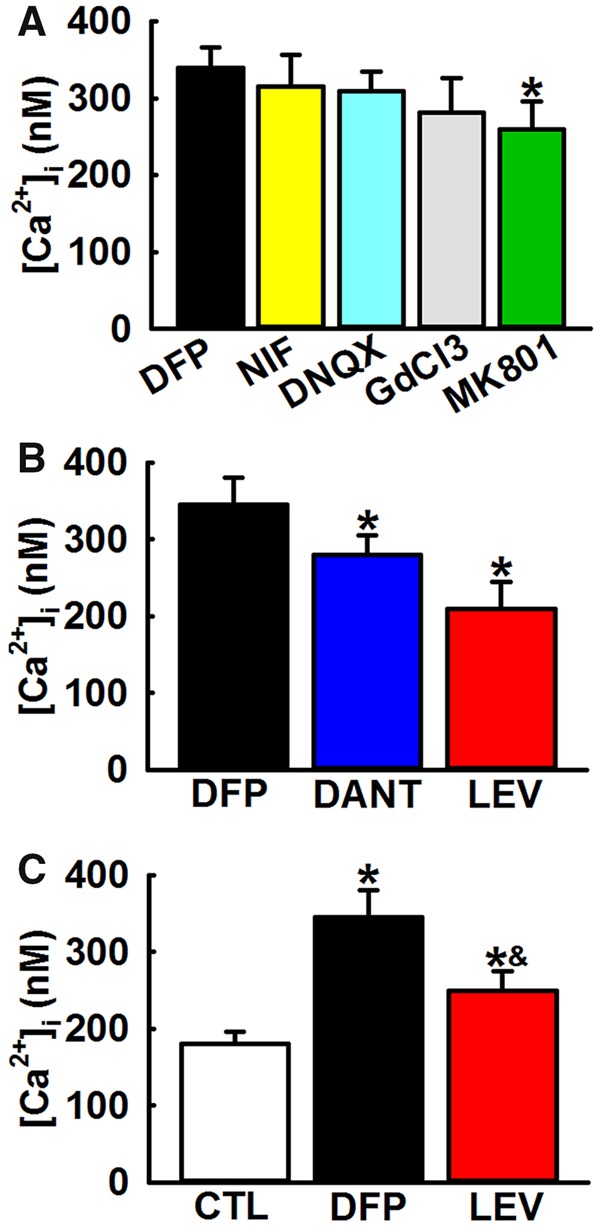
Mechanism for elevated hippocampal [Ca2+]i in DFP rats. A, Effect of inhibitors of Ca2+ influx pathways on [Ca2+]i in DFP. Bath application of inhibitors of voltage-gated Ca2+ channels (Nifedipine, 5 μM), AMPA/kainate channels (DNQX, 10 μM) or other nonspecific cation channels (GdCl3, 10 μM) did not produce a significant decrease in [Ca2+]i. Bath application of NMDA antagonist (MK-801, 10 μM) produced a small but significant reduction in [Ca2+]i (Data expressed as mean ± SEM, *p < .05, t test, n = 5 rats). B, Effect of inhibitors of intracellular Ca2+ release pathways on [Ca2+]i in DFP neurons. Bath application of IP3/RyR antagonist LEV (100 μM) or RyR antagonist dantrolene (Dant, 50 μM) produced significant reductions in [Ca2+]i. (Data expressed as mean ± SEM, *p < .05, t test, n = 6 rats). C, Effect of LEV administration on [Ca2+]i in DFP rats. Treatment with LEV (50 mg/kg, i.p.) produced a significant reduction in [Ca2+]i. compared with DFP neurons but was significantly higher than age-matched control neurons (CTL) (Data expressed as mean ± SEM, *p < .05, 1-way ANOVA, Tukey’s test, n = 5 rats).
